Mechanisms for initiating cellular DNA replication Biology Diagrams
Mechanisms for initiating cellular DNA replication Biology Diagrams DNA replication timing varies among individuals at specific loci. A. FACS-sorting cells by DNA content enables analysis of DNA copy number (by whole-genome sequencing) in G1 and S phase cells (adapted from Koren et al., 2012).. B. Analysis of the ratio of DNA copy number between S- and G1-phase cells along each chromosome allows the construction of replication timing profiles; early

The study of genome architecture and DNA replication timing in cleavage-stage embryos will be facilitated by the recent development of single-cell Hi-C, single-cell RNA sequencing and single-cell DNA replication timing is deterministic at the level of chromosomal domains but stochastic at the level of replicons in Xenopus egg extracts. Nucleic Acids Res 36: 5623-5634 [PMC free article] [Google Scholar] Lande-Diner L, Zhang J, Cedar H 2009. Shifts in replication timing actively affect histone acetylation during nucleosome reassembly.
DNA replication timing: random thoughts about origin firing Biology Diagrams
Aberrant DNA replication timing is associated with changes in gene expression, changes in epigenetic modifications and an increased frequency of structural rearrangements. Furthermore, certain replication timing changes can directly lead to overt genomic instability and may explain unique mutational signatures that are present in cells that DNA replication timing influences gene expression level. J Cell Biol. 2017; 216:1907-1914. Whole genome replication timing analysis of 7 phylogenetically diverse yeast identified genomic features with conserved replication timing and demonstrated that early replication timing might facilitate maximal expression of genes expressed in S-phase

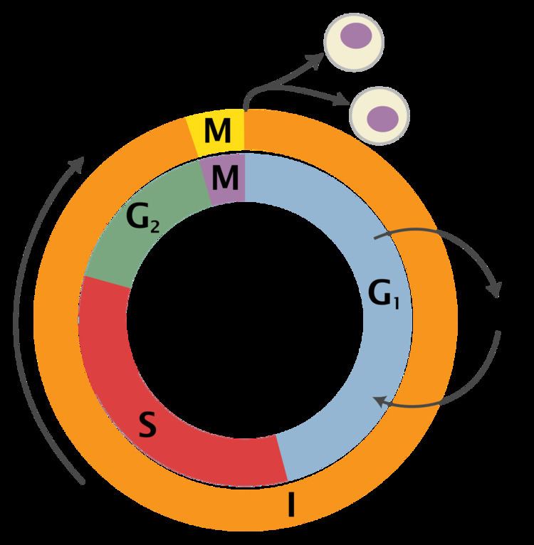
Keywords: DNA replication timing, DNA replication origin, S-phase regulation, MCM, ORC, Stochastic model, Origin activation. Introduction. The temporal organization of DNA replication during S phase is a fascinating subject on many levels.

Genetic variation in human DNA replication timing Biology Diagrams
Figure 4: A diagrammatic representation of replication timing in a 70-Mb segment of human chromosome 2.The red horizontal line represents time in S-phase, from early (top) to late (bottom). Grey data points each represent a different DNA sequence position along the length of chromosome 2 as indicated on the x axis, with more positive values on the y-axis indicating earlier replication. DNA replication timing leads to variation in DNA copy number along chromosomes among S phase cells (e.g., early-replicating regions are duplicated in most cells), causing read depth fluctuations
